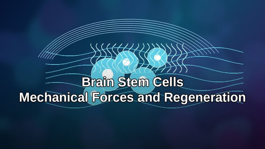
Shiri Gur-Cohen, Ph.D., UC San Diego, explains how her lab studies how epithelial stem cells communicate with their vascular microenvironment to advance regeneration and combat metastatic diseases.
Video Transcript:

[MUSIC] I’m excited to share with you today the work that we’re doing in my laboratory to enhance regeneration and to fight cancer. In my laboratory, we study how stem cells communicate with their environment. By doing that, I want to focus today on how stem cells can reconstruct their lymphatic niche during regeneration and what happen in cancer in aging. Let me start by telling you that our body has a remarkable regenerative capacity. In a single day we lose and replace billions of cells and that’s completely normal.
The way that I like to think about it is that we all know how we woke up this morning, but we must have changed several times since then. This is not just a fantasy story out of Alice in Wonderland. There’s also a paradox in that because we know that a key players in this regenerative process are the stem cells and they have a balanced cell fate decision. They know when and where to create a tissue. But stem cells can also age.
If they are creating less tissue, we’re aging, but if they do too much of what they need to do, we can develop diseases such as cancer.
Essentially, we’re missing a fundamental understanding of what dictates stem cell fate decision and how this is balanced throughout regeneration. The skin is really an excellent and exciting model to ask these type of questions because the skin is a house for multiple types of stem cells, including those that create the skin on your body that the barrier from the external environment and the hair follicles to grow. This is the beautiful picture that you see in the screen. Essentially what we wanted to understand is how the stem cells that exist in the skin can integrate systemic changes across them to balance their fate cell decisions.
One system that potentiate to regulate that process is the vascular system. They create a network in our body that can really synchronize those signals that pass through our body to the stem cells. Normally when I talk about the vascular system, most of you probably think about the blood vessels that carry cells and nutrients into our tissues. But we also have one more system and this is the lymphatic vascular system. This system will drain excess fluids and macromolecules from the tissue back into the circulatory system.
Now I can really focus and ask how the vascular system coordinate the regenerative process in the skin. To do that, we use three-dimensional imaging and tissue clearing. What you can see here in red are the lymphatic capillaries into skin. We were pretty much amazed to see how the lymphatic capillaries forming a niche for the stem cells in the hair follicle stem cells. We could also model that type of interaction and to visualize how the stem cells are nesting on top of the lymphatic capillaries when they’re at the resting phase.
This is when the stem cells are basically not doing anything. They are existing in the skin and they’re not growing hair at that point. Now we’re going to take a virtual tour into our skin to better understand the nature of those interactions.
What we noticed is that the lymphatic capillaries, here in white, are creating protrusions that emerge towards the stem cells and holding them together protecting them, doing something to them to make them quiescence or basically in the resting phase. We wanted to understand why is that happening and why is that important.
We started by profiling the stem cells themselves. When we profiled them using either bulk RNA sequencing or single-cell RNA sequencing, we found something very remarkable. We found that the stem cells themselves highly expressive protein called angiopoietin-like 7 that is known to regulate vascular biology and lymphatic biology.
But it was only in the stem cells and not in any other cell type in the skin. You can also see it very clearly in the single-cell RNA sequencing.
The stem cells themselves secrete lymph angiogenic factor. Essentially what we found is that the stem cells themselves in the hair follicle secrete angiopoietin-like 7 to control how lymphatic will drain fluids and macromolecules. On the other hand, when the stem cells turn into their regenerative state, then they secrete other factor. They are switching their secretum to secrete angiopoietin-like 4 and that control reduce lymphatic drainage and an ability to control how stem cell fate decision will look like, whether they will regenerate a tissue or will not regenerate a tissue.
Essentially what we found is that we have a new niche cell component right now in hand that nobody ever heard before.
This is the lymphatic vessels, and that the stem cells themselves can shape their lymphatic microenvironment. But it’s true to all of our tissues because what I showed you so far is that this is true for the hair follicle stem cells, but these stem cells act in cycles.
They will either grow or don’t grow hair. This is why we see cycles of hair growth on our body. But some other organs such as the intestine, is constantly regenerating all the time.
Those stem cells in the intestine are constantly activated. Can it be that one niche like the lymphatic microenvironment rule or dictate cell fate decisions in the intestine, in the skin in a very similar way even though their regenerative demand is so different? This is actually stemming from a very fun and important collaboration between gastroenterologist in Waill Cornell and computational genius in the lab of Dana Pe’er, where we were trying to understand how lymphatics in the intestine is working in forming a niche for the intestinal stem cells. We first started to profile the vascular system in the intestine. In red you can see the blood vessels and as expected, the intestine is highly vascularized.
But we could also see that there is a very rich vascular network of lymphatic capillaries that reside just beneath the crypt where the stems are residing. Using, again, three-dimensional imaging, we could visualize and see how the LGR5 positive stem cells in the intestine, here in green, are nesting on top of the lymphatic capillaries also in the intestine, and that is the happening across the intestine. No matter if it was the colon or the small intestine, that type of interaction was consistent all along. Why is that important here? Why does stem cells here need the lymphatics?
To understand that we established a co-culture system where we can take the organoids that are forming in the intestine in vitro and to grow them on top of lymphatic capillaries.
The beauties that in culture those intestinal stem cells can form the crypt base and to differentiate so we can measure or trace how their balanced cell fate decisions are happening. What we saw is that when the stem cells are being cultured on top of lymphatic capillaries, the stem cells are maintaining their identity, but they’re not doing much. They cannot really regenerate, they cannot produce the differentiated progeny. How is that happening?
Why is that happening? To do that, we turned into spatial transcriptomics to understand how the lymphatics at the base of the crypt can produce factors or do something that is very different from lymphatics that are not nesting the stem cells like those in the villus. We performed spatial transcriptomics and we projected the data on the crypt-villi axis.
This is the data that you can see here and we could beautifully see lymphatics. This is the purple line over here.
The lymphatics that are nesting at the base versus lymphatic that are at the villus. Now we can ask, what is the difference between lymphatics at the base versus those that are at the villus and are not touching stem cells? What we found is that the lymphatic at the base are actually secreting factors that are considered to be stem cell factor, known stem cell factor like R-spondin-3. But we also found a novel factor called RELN. RELN was specifically expressed at the crypt base but not in the villi and we found by functional data that this RELN can dictate how cell fate decisions are happening, what basically cause the stem cells to either produce differentiated cells or not produce differentiated cells.
What I showed you right now is that we have the lymphatic niche that again, this is completely new to our knowledge, but it will form a niche to different tissues with completely distinct regenerative domains.
But we also all know that we find a niche that is important for regeneration but we all know the regeneration is an arrow with a one negative direction and aging will affect or affecting all of us. We’re spending a lot of time in the lab thinking how we can treat the stem cells, how we can manipulate the stem cells, or target the lymphatic stem cell interaction so we can maintain this regenerative capacity. Or more importantly, we can also think about how we can reverse aging, how we can take those stem cells that are considered to be aged or the environment that is considered to be aged and to engineer the system differently. As I said, we’re all going to experience aging.
It’s not something that is completely far away from us. How do you know that you age? The skin is usually the first marker for us being aging.
During aging, the skin texture is different, we see more wrinkles. The wounds on our skin take so much more time to heal than we were young.
How is the microenvironment or the lymphatic micro-environment look like in the aged skin? That’s a question that we were interested in. What we found is that when we age, lymphatics no longer maintain their structure, they become dilated, they’re dysfunctional and they’re no longer, you can clearly see that their structure is completely different. But what’s happening to the association of stem cells with lymphatics? We know that the structure is different, but are they still associated with lymphatics?
This is exactly what we did. Again, using three-dimensional imaging, you can see here the stem cells in yellow.
But we were amazed to see that the blood vessels that were considered to be the factor of aging are still highly associated with the stem cells, but lymphatics are no longer during aging. That was very surprising to us. When we looked at mice that we engineered to have a dysfunctional lymphatics, they presented a phenotype of premature hair loss.
If you don’t have functional lymphatics, those hair follicle stem cells will no longer regenerate the tissue as much as they need to regenerate. Thinking forward, we know association right now, we know that excessive or insufficient lymphatic function can give rise to a variety of pathologies or regenerative pathologies including delayed wound repair. I showed you that it’s also associated with aging. Later on the road, we know the cancer cells are using lymphatics as one of the major exit routes from the primary tumor to seed metastatic diseases in distant organs.
What we want to do is to target the lymphatic stem cell interaction so we can enhance regeneration, but at the same time we want to fight cancer and we want to fight the ability of those cancer stem cells to disseminate through lymphatics.
How do we do it? How do we start to target that system? We engineered mice that will have disrupted and you’re putting seven expression, this factor that is expressed by the stem cells. What we found is that again, when we are not having these lymphangiogenic factors that is secreted by the stem cells, lymphatics are no longer intact and they’re massively dilated.
How the stem cell look like?
What we found is that in these mice that have dysfunctional under pointing like seven secretion specifically by the stem cells, the hair follicle stem cells started to develop hyperplasia. They became big. Their balanced cell fate decisions that we started to talk about were no longer there, you can see those big follicles.
But these big follicles that you can see here, we wanted to understand their association with lymphatics. What we found is that they were highly associated with an emphatic capillaries and those capillaries that you see here are humongous.
They are big, very big lymphatics that are surrounding the hyperplastic hair follicle stem cells. When we looked closer in completely different mouse model that have dysfunctional lymphatics, we found similar things that happening within the stem cells. We saw that they’re starting to accumulate lots of DNA damage in the form of gamma-H2AX, which you can see here in magenta. It didn’t matter if it was acute lymphatic disruption or if it was chronic lymphatic disruption, both models show that stem cells starts to accumulate lots of DNA damage. Why is that important?
Because with time, especially in our skin, if we accumulate lots of DNA damage, we can develop cancer and this is one of the forms that cancer can be developed and is squamous cell carcinoma of the skin and this is one of the most common form of skin cancer.
It accounts for about 1.8 million patients a year in the United States only. It’s a lot of patients. Although it rarely metastasize, when it does, it present a very poor prognosis with a very low survival rate over 10 years.
We need a better solution to fight cancer. In skin cancer, at least at the heart of those tumors they’re starting to develop, we have stem cells that will fuel the growth of the tumor, but those stem cells are not working in a void. They have environment that will support them. If we again want to fight cancer, we want to understand how the cancer stem cells are making their environment to work for them or the healthy environment to work for them so they can thrive and metastasize at the end. Thinking forward, we wanted to understand how those tumor stem cells can interact with their lymphatic microenvironment.
We looked at the mouse model that have squamous cell carcinoma and this mouse model cells or the stem cells were marked with a reporter, and this is what you can see here. Those are red cells. Those are the stem cells within the squamous cell carcinoma of the skin.
What we found is when the tumor develop, it starts to be highly associated also with lymphatic capillaries. Right now we have a project that is running in the lab to understand how the tumor cell of origin and how the stem cells that create this tumor form and fuel the growth of the tumor interacting with lymphatics and whether we can target that interaction so we can prevent tumors to use lymphatics as the exit route in seed metastases.
Just to wrap up, my laboratory really study how epithelial stem cells communicate with their microenvironment. With the idea that we want to advance tissue regeneration, we want to change the way that we age, we want to change the way that we heal wounds and eventually we want to combat also metastatic diseases. With that, we also have positions. We have really vibrant group that is already started to emerge. We have exciting projects.
If you know anyone that is interested, please spread the word. With that, I want to thank my amazing lab members already there helping me so much to establish a lab which is, it’s a hard work and they’re doing it together with me and it’s really fun.
My collaborators across the world, my funding resources that help to support the work that I’ve showed you today. Thank you so much for your attention. I think we do have time for a few questions, if they have the questions to you.
Anyone have questions? Yes, Kate. We talked about this before and now hold throughout, I had brought up these Andrew pointings are actually unusual in their structure, may be difficult to target. How are you thinking about and you put it in like seven and four in terms of restoring the balance? Yeah.
It’s a good question. I think in general this is the Andrew pointing like family, if we think about them they are new to science. We don’t know much about their structure.
We don’t know almost anything about their receptors. Scientifically, I think it’s really hard for us to try to target it, but we are developing new tools and technologies that enables us now to crisper out or knock in those factors so we can manipulate their secretion.
What we basically do, we are going to change or target the stems are secret on how the stem cells are taking advantage of their environment to make regeneration so we can do it ourselves. We’re developing those tools, were developing those technologies. But I think for your question also, we want to understand the mechanism and this is very important for us to understand the mechanism because the Andrew pointing like seven and four maybe right in the skin, but not in other organs, for example, but if we understand the mechanism of how we manipulate lymphatic, what lymphatics needs to, to control stem cell behavior, then we can bypass this product and do it.
These are things that we’re constantly thinking of doing in the lab. Intro lymphatic delivery of anticancer drugs for skin cancer.
Is that something that’s people are looking at? Intro lymphatic, can you please repeat again? Intro lymphatic delivery. Intro lymphatic delivery. Anticancer drugs.
Great question, I think in general, what exists in the lymphatic start? Well, what’s inside of that? If you just look at what exists inside so we can target it or inject something into it. The traditional view is that what exists inside lymphatics is a trash. The emphatic is like the garbage trash off the body, absorb all the fluids, macromolecules that we don’t need and it’s important for immune surveillance.
All the immune cells that are, I’m eating all of those antigens. We’ll go through lymphatics and educate themselves in the lymph node. Thinking about it, what exists there, is there at all the French are distinct drainage ability so we can control what’s going into lymphatic? Maybe we can target for example, fat. We know that in the intestine, lymphatics are important, at least the villis to absorb fat and there have been reports that it’s important to inhibit obesity.
I think if we would understand better how distinct kind of regulation of drainage is happening. Maybe we can target drainage and maybe we can inject specific molecules into the lymphatics. They can travel through the body because one of the things that I haven’t shown in this work is that when we manipulate the lymphatics to become dysfunctional, the stem cells will no longer be balanced, but also synchronize regeneration will not happen. One stem cells will no longer know about the other stem cells they fit, needs to regenerate a tissue or not. Synchronous or regeneration is an important thing.
For your question, can we actually inject something into the lymphatic to synchronized tissue regeneration, not only the skin but maybe in the intestine, in the lung.
To synchronize this regenerative process is something that hasn’t been worked out at all. Yes. Hi, great talk. Thank you.
I actually want to expand on something you said for synchronized regeneration. Have you ever walked into cellular senescence with regards to all of this? Because there has been mounting evidence, like in the camp PC lab and the Buck Institute that the influx of cellular senescence is actually needed for synchronized regeneration of skin tissue. There’s also been a study in skeletal muscle where they did a single cell RNA seek, and most senescent cells are actually foreign immune cells infiltrating into the tissue to help repair it. I’m wondering if you’ve ever looked into that to see the underlying mechanism, if there was any interaction with these immune cells are expressing senescent markers and in the lymphatic vessels.
I love this question because I think in general we were talking on mechanism and how is that happening? Senescence may be one of the part of what exactly legal fight it goes doing there to the stem cells. One of the things that I find really fascinating is that it’s a network, it’s not only a mesenchymal cells that will nester stem cells. It’s a network, they will exist across the tissue. If you think about the intestine, the lungs, or the skin, we have this as a system, as a network that can sample things and can deliver information from one site to another.
You’re talking about senescence. In the skin that hasn’t been worked out at all, but at least in the intestine, there’s something very interesting because if you induce an essence in one spot, the other distance spot will sense it and becomes senescence as well and nobody knows why. I think it’s really interesting way to think about in general, synchronized regeneration, synchronized aging. If we can target it to synchronized regeneration across the tissues. To your question, I haven’t looked at senescence, but I love this question.
[LAUGHTER] [MUSIC]