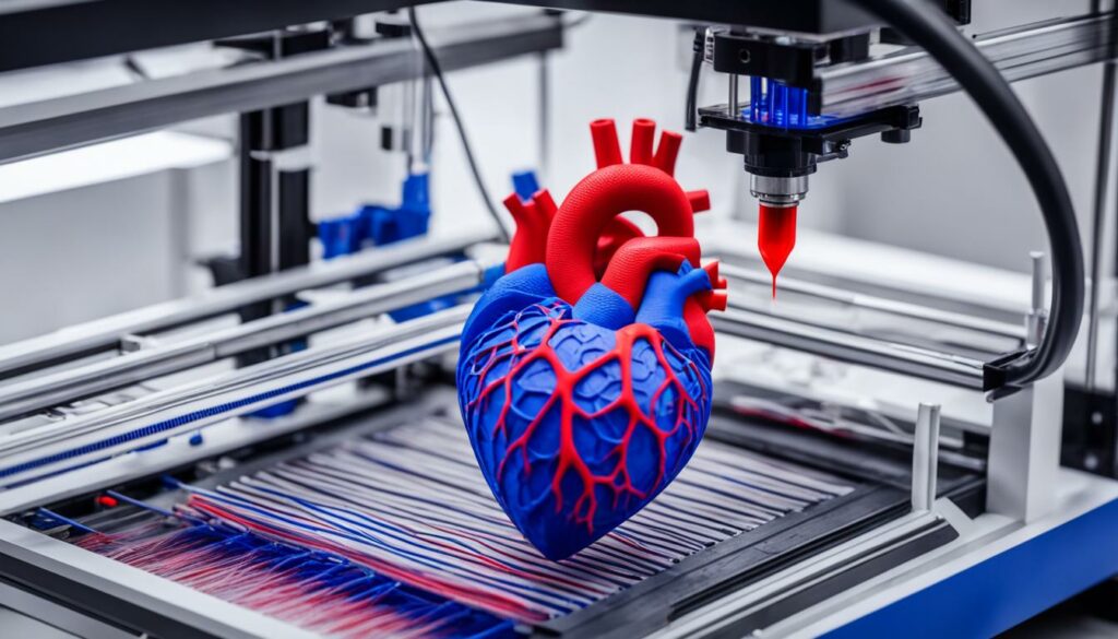
We are witnessing remarkable advancements in the field of 3D bioprinting that are revolutionizing cardiovascular disorder treatment and improving heart care. This cutting-edge technology combines engineering, biology, and medicine to create precise and functional structures that can aid in the regeneration and repair of cardiac tissue.
Key Takeaways:
- 3D bioprinting is transforming the field of cardiovascular disorder treatment.
- Cardiac tissue engineering aims to repair damaged or diseased cardiac tissue.
- Various bioprinting techniques and bioinks are used to create complex 3D structures.
- Applications of 3D bioprinting in cardiac tissue engineering include heart valve repair and vascularized tissue creation.
- Ongoing advancements in bioprinting techniques are improving the precision and efficiency of cardiac tissue engineering.
Understanding Cardiac Tissue Engineering
Cardiac tissue engineering is a fascinating field that combines principles from materials engineering, life sciences, and computer modeling to develop innovative strategies for regenerating damaged or diseased heart tissue. Through the creation of functional tissue equivalents, researchers aim to restore the functionality and structure of the heart, offering new hope for patients with cardiovascular disorders.
At the core of cardiac tissue engineering lies the concept of tissue engineering itself. Tissue engineering focuses on the development of biological substitutes that can repair or replace damaged tissues or organs. By harnessing the potential of biomaterials, cells, and growth factors, tissue engineers aim to mimic the native environment of the targeted tissue and facilitate its regeneration.
In the context of cardiac tissue engineering, the goals are specific to the heart and its intricate structure. The intricate nature and complex functionality of cardiac tissue present unique challenges that require innovative approaches. Through meticulous laboratory research and multidisciplinary collaboration, scientists are striving to understand and recreate the complexity of the heart.
The ultimate aim of cardiac tissue engineering is to develop strategies that can effectively regenerate cardiac tissue and restore its function. This can be achieved through the creation of three-dimensional structures that closely resemble the native cardiac tissue. By mimicking the organization of cells and extracellular matrix in the heart, tissue engineers hope to promote cell survival, integration, and function in the engineered tissues.
One promising approach is the use of biomaterials as scaffolds to support and guide the growth of cells, allowing them to organize and function as they would in a healthy heart. These biomaterials can be engineered to provide mechanical support, deliver bioactive molecules, and facilitate vascularization, all crucial components for the successful regeneration of cardiac tissue.
Cardiac tissue engineering is a multidisciplinary field that combines principles from materials engineering, life sciences, and computer modeling to develop strategies for regenerating damaged or diseased heart tissue.
The dynamic nature of cardiac tissue engineering research has yielded significant advancements in recent years. By understanding the biology of the heart and leveraging innovative technologies, researchers are making great strides in the quest for heart regeneration. From creating bioprinted cardiac constructs to developing functional cardiac patches, the potential of cardiac tissue engineering is truly transformative.
As we continue to explore and unravel the intricacies of cardiac tissue engineering, the possibilities for cardiovascular disorder treatment and heart regeneration become increasingly promising. The synergistic integration of biology, materials science, and engineering provides a solid foundation for overcoming existing challenges and driving innovative breakthroughs in the field. Through ongoing research and collaboration, we are committed to advancing the frontiers of cardiac tissue engineering and improving the lives of patients with heart-related disorders.
The Basics of 3D Bioprinting
3D bioprinting is a transformative technology that allows us to fabricate complex 3D structures by depositing layers of biomaterials. This groundbreaking technique has opened up new possibilities in various fields, including medicine, regenerative biotechnology, and tissue engineering.
Bioprinting techniques are at the core of this technology, enabling precise control over the deposition of biomaterials to create intricate structures. Let’s explore some of the most commonly used bioprinting techniques:
- Droplet-based bioprinting: In this technique, droplets of bio-ink are dispensed and carefully positioned to build up the desired structure layer by layer. This method offers high resolution and allows for the deposition of multiple cell types within a single structure.
- Laser-assisted bioprinting: Laser pulses are used to propel bio-ink from a donor slide to a collector substrate, creating a precise pattern. This technique is particularly useful for printing delicate structures or cells with high viability requirements.
- Stereolithography-based bioprinting: Stereolithography utilizes a beam of light to selectively cure photopolymerizable bio-inks, layer by layer, to create complex 3D structures. This method offers high resolution and can produce intricate constructs with varying mechanical properties.
- Extrusion-based bioprinting: In this widely used technique, bio-ink is extruded through a nozzle or syringe to create the desired structure. The extrusion process can be controlled to deposit cells, biomaterials, and other additives simultaneously, allowing for the creation of complex tissues.
The choice of bioprinting technique depends on the specific requirements of the desired structure and the biomaterials used. Each technique has its advantages and limitations, and researchers continue to explore and refine these techniques to expand their capabilities.
Bioprinting is a multidisciplinary field that encompasses not only the printing techniques but also the biomaterials used. These biomaterials, commonly known as bio-inks, serve as the building blocks for printing functional tissues and organs.
Biomaterials used in 3D bioprinting should possess certain characteristics, such as biocompatibility, biodegradability, and the ability to support cellular growth and differentiation. Commonly used biomaterials include natural polymers like alginate, collagen, and fibrin, as well as synthetic polymers and hydrogels.
By combining the various bioprinting techniques and biomaterials, researchers are pushing the boundaries of 3D bioprinting, paving the way for personalized medicine, tissue engineering, and regenerative therapies.
Applications of 3D Bioprinting in Cardiac Tissue Engineering
3D bioprinting has opened up remarkable possibilities in the field of cardiac tissue engineering, revolutionizing heart valve repair and regeneration strategies. Through this groundbreaking technology, bioprinted cardiac constructs, including heart valves, vascularized tissue, and functional cardiac patches, have been successfully created. These advancements hold tremendous potential for treating cardiovascular disorders and improving heart health.
In cardiac tissue engineering, the creation of 3D bioprinted constructs has provided a precise and customizable approach to repair damaged heart tissue. Bioprinted heart valves offer an alternative to traditional prosthetic valves, as they can be tailored to the patient’s specific anatomy and exhibit better compatibility and functionality. This personalized approach enhances the success of heart valve replacements and reduces the risk of complications.
| Applications of 3D Bioprinting in Cardiac Tissue Engineering | Advantages |
|---|---|
| Heart valve repair and replacement | – Enhanced compatibility and functionality – Personalized anatomical fit |
| Vascularized tissue creation | – Improved blood circulation in bioprinted constructs – Enhanced tissue viability and functionality |
| Functional cardiac patches | – Regeneration of damaged cardiac tissue – Improved heart function and contractility |
Another significant application of 3D bioprinting in cardiac tissue engineering is the creation of vascularized tissue. By incorporating intricate vascular networks within bioprinted constructs, researchers aim to enhance blood circulation and provide nutrients to the engineered cardiac tissue. This approach offers improved tissue viability and functionality, which is crucial for successful tissue integration in transplantation and regenerative medicine.
Functional cardiac patches, produced through 3D bioprinting, hold great potential for repairing damaged or scarred cardiac tissue. These patches can be designed to mimic the native heart tissue, promoting cell growth, maturation, and contractility. The bioprinted cardiac patches facilitate the regeneration of damaged tissue and aid in restoring the heart’s normal function.
Overall, the applications of 3D bioprinting in cardiac tissue engineering have significantly advanced the field, offering tailored and functional solutions for heart valve repair, vascularized tissue creation, and cardiac patch development. As research and technology continue to evolve, these advancements hold immense promise for the future of cardiovascular disorder treatment.
Advancements in Bioprinting Techniques for Cardiac Tissue Engineering
Bioprinting techniques play a crucial role in the field of cardiac tissue engineering, enabling the creation of complex and functional cardiac tissue constructs. These techniques have undergone significant advancements, enhancing precision, efficiency, and the overall quality of the bioprinted tissues.
One of the notable bioprinting techniques is droplet-based bioprinting, which involves the precise deposition of cell-laden bio-ink droplets to form intricate structures. This technique has seen remarkable progress in terms of improved cell viability, higher resolution, and enhanced printability, making it an excellent choice for creating fine and intricate cardiac tissue models.

Another notable technique is extrusion-based bioprinting, which uses a syringe-based system to extrude bio-ink through a nozzle. This technique offers excellent control over the deposition process, allowing for the creation of large-scale, anatomically accurate cardiac tissue constructs. Recent advancements in extrusion-based bioprinting have focused on improving the resolution and cell viability, resulting in more reliable and functional bioprinted cardiac tissues.
“Bioprinting techniques, such as droplet-based and extrusion-based bioprinting, have played a significant role in advancing cardiovascular tissue engineering. These techniques have improved the precision and efficiency of creating cardiac tissue constructs with enhanced functionality.”
The third notable technique is inkjet bioprinting, which employs piezoelectric technology to deposit bio-ink droplets onto a substrate. Inkjet bioprinting offers high-resolution printing capabilities with precise control over droplet size and placement. Recent advancements in inkjet bioprinting have focused on improving cell viability and enhancing the printability of various bio-inks, enabling the creation of complex and functional cardiac tissue models.
The Advancements in Bioprinting Techniques:
- Improved cell viability
- Higher resolution
- Enhanced printability
| Bioprinting Technique | Advancements |
|---|---|
| Droplet-based bioprinting | Improved cell viability, higher resolution, enhanced printability |
| Extrusion-based bioprinting | Enhanced resolution, improved cell viability |
| Inkjet bioprinting | Improved cell viability, enhanced printability |
These advancements in bioprinting techniques have significantly contributed to the progress in cardiac tissue engineering. The improved precision, resolution, and cell viability provided by droplet-based, extrusion-based, and inkjet bioprinting techniques have paved the way for the creation of functional and anatomically accurate cardiac tissue constructs. As these techniques continue to evolve, the prospects for cardiac tissue engineering and cardiovascular disorder treatment become even more promising.
Bioinks for 3D Bioprinting
In the field of 3D bioprinting, bioinks play a crucial role in creating the intricate structures needed for tissue engineering. These materials serve as the “ink” that is deposited layer by layer to build complex three-dimensional constructs. One of the most commonly used types of bioinks is hydrogels, which offer remarkable biocompatibility and the ability to replicate the natural extracellular matrix (ECM).
Hydrogels are composed of water-rich networks, making them an ideal choice for supporting cell growth, differentiation, and tissue regeneration. Within the hydrogel category, several biomaterials have gained prominence in 3D bioprinting:
- Alginate: Alginate is a natural polysaccharide derived from brown seaweed. Its gel-like consistency and biocompatibility make it an excellent choice for creating bioinks. Alginate-based bioinks offer good printability and biodegradability, making them suitable for various tissue engineering applications.
- Collagen: Collagen is the most abundant protein in the human body and is a vital component of connective tissues. As a bioink, collagen enables cell adhesion and supports the formation of tissue structures. Its biodegradability and biocompatibility make it highly desirable for tissue regeneration purposes.
- Fibrin: Fibrin is a fibrous protein involved in blood clotting and wound healing. It can be transformed into a gel-based bioink by enzymatic crosslinking. Fibrin bioinks provide an environment conducive to cell migration, proliferation, and the formation of vascular networks, making them particularly suitable for cardiac tissue engineering.
These bioinks can be further modified by incorporating growth factors, cytokines, and other bioactive molecules to enhance their functionality and influence cell behavior. This customization allows researchers to mimic the native tissue microenvironment more accurately and achieve better tissue integration and regeneration.
“The use of bioinks, particularly hydrogels, has revolutionized the field of 3D bioprinting. By carefully selecting and formulating bioinks, we can create a biomimetic environment that promotes tissue regeneration and ultimately improves patient outcomes.” – Dr. Emily Thompson, Bioprinting Researcher
3D bioprinting, combined with these advanced bioinks, has opened up exciting possibilities for creating patient-specific tissues and organs. Through precise control of the printing process and the incorporation of bioactive components, researchers can develop complex structures that closely resemble native tissue architecture.
To give you a better idea of the potential of bioinks, take a look at the following visual representation:
| Bioink | Main Characteristics | Applications |
|---|---|---|
| Alginate | Good printability, biocompatibility, biodegradability | Organoids, skin tissue, cartilage engineering |
| Collagen | Cell adhesion, biocompatibility, biomimicry | Skin tissue, wound healing, nerve regeneration |
| Fibrin | Promotes cell migration, vascularization, natural clot formation | Cardiac tissue, blood vessels, wound healing |
Researchers continue to explore and optimize various bioinks to broaden their applications and enhance their performance. Advances in bioink formulations, printing techniques, and tissue engineering approaches are paving the way for the development of functional and viable bioprinted tissues and organs.
With the rapid progress in the field of 3D bioprinting, we are moving closer to the day when bioprinted tissues and organs can help alleviate the shortage of donor organs and provide personalized solutions for patients with cardiovascular disorders and other medical conditions.
Challenges and Limitations of 3D Bioprinting for Cardiovascular Disorder Treatment
While 3D bioprinting holds great potential for cardiovascular disorder treatment, there are still several challenges and limitations that need to be addressed. These include the need for further improvements in cell viability, vascularization, scalability, and regulatory approval. We understand the importance of overcoming these challenges to ensure the widespread clinical application of 3D bioprinting in cardiac tissue engineering.
One of the key challenges of 3D bioprinting is achieving high cell viability in the printed structures. The process of bioprinting can subject cells to shear stress and temperature changes, which can affect their viability and functionality. It is crucial to optimize bioprinting parameters, such as printing speed, nozzle diameter, and pressure, to minimize cell damage and ensure successful tissue integration.
Vascularization is another critical aspect that needs to be addressed. To replicate the complex network of blood vessels in cardiac tissue, researchers need to develop techniques for printing functional vascular structures. Without a sufficient blood supply, bioprinted cardiac tissue may not survive or function properly. Strategies such as bioprinting prevascularized constructs or incorporating vasculature-inducing factors can help overcome this limitation.
Scalability is another challenge in 3D bioprinting. While researchers have successfully printed small-scale cardiac tissue constructs, achieving larger, clinically relevant sizes remains a challenge. The scalability of bioprinting processes and the availability of bioreactors capable of supporting the growth and maturation of large bioprinted tissues are areas that require further development. We are actively exploring innovative approaches to scale up bioprinting techniques and ensure the production of functional tissue constructs at a clinically relevant scale.
Regulatory approval is also an important consideration in the advancement of 3D bioprinting for cardiovascular disorder treatment. The bioprinted tissues and constructs need to meet rigorous safety and efficacy standards set by regulatory authorities before they can be used in clinical settings. Compliance with these regulations ensures the reliability and effectiveness of the bioprinted treatments. We are committed to working closely with regulatory agencies to meet these standards and bring 3D bioprinting innovations to the patients who need them.
Challenges and Limitations of 3D Bioprinting in Cardiovascular Disorder Treatment
| Challenges | Solutions |
|---|---|
| Cell viability | Optimizing bioprinting parameters to minimize cell damage |
| Vascularization | Developing techniques for printing functional vascular structures |
| Scalability | Advancing bioprinting processes and bioreactor technologies for larger tissue constructs |
| Regulatory approval | Meeting safety and efficacy standards set by regulatory authorities |
Addressing these challenges will be crucial for harnessing the full potential of 3D bioprinting in cardiovascular disorder treatment. Despite these limitations, we remain optimistic about the future advancements in this field and are committed to overcoming these challenges to improve heart health and revolutionize the field of cardiovascular disorder treatment.
Future Directions for 3D Bioprinting in Cardiac Tissue Engineering
The future of 3D bioprinting in cardiac tissue engineering holds immense potential for transforming regenerative medicine. Researchers are actively exploring innovative approaches and technologies to enhance the functionality and maturation of bioprinted cardiac tissue. These advancements are paving the way for the development of patient-specific treatments and revolutionizing the field of cardiovascular disorder treatment.
Exploring New Bioinks
One of the key areas of focus in future research is the development of advanced bioinks for 3D bioprinting. Scientists are investigating the use of decellularized extracellular matrix (ECM) as a bioink, which can provide a more native and supportive environment for cell growth and tissue regeneration. This approach aims to improve the structural integrity and functionality of the bioprinted cardiac tissue, bringing it closer to its natural counterpart.
Incorporating Advanced Technologies
To overcome the current limitations of 3D bioprinting in cardiac tissue engineering, researchers are integrating other cutting-edge technologies into the process. Bioreactors, for example, can provide controlled environments to enhance cell viability, nutrient supply, and tissue maturation. Organ-on-a-chip systems, on the other hand, enable the replication of complex physiological conditions, allowing for more realistic testing and analysis of bioprinted cardiac tissue.
“The integration of advanced bioprinting technologies with other innovative approaches holds tremendous promise for the future of cardiac tissue engineering.”
Advancing Patient-Specific Treatments
With the advancements in 3D bioprinting, the potential for creating patient-specific treatments is becoming a reality. By utilizing patient-specific cells and imaging techniques, scientists can design and bioprint customized cardiac tissue constructs tailored to each individual. This personalized approach has the potential to improve treatment outcomes and reduce the risk of rejection, ultimately leading to more successful heart care.
Developing Functional Vascular Networks
Another crucial aspect of future research in cardiac tissue engineering is the development of functional vascular networks within bioprinted constructs. The establishment of blood vessel networks is essential for supplying oxygen, nutrients, and removing waste products from the tissue. Scientists are working towards creating intricate and efficient vascular networks that can seamlessly integrate with the bioprinted cardiac tissue, promoting its survival and functionality.
To summarize, the future of 3D bioprinting in cardiac tissue engineering holds great promise for regenerative medicine. Through the exploration of new bioinks, incorporation of advanced technologies, development of patient-specific treatments, and the creation of functional vascular networks, researchers are pushing the boundaries of what is possible in the field. These future directions will undoubtedly contribute to significant advancements in cardiac tissue engineering and revolutionize the treatment of cardiovascular disorders.
Conclusion
The field of 3D bioprinting has witnessed rapid advancements, ushering in new possibilities for cardiac tissue engineering and the treatment of cardiovascular disorders. Through the integration of innovative bioprinting techniques, bioinks, and tissue engineering approaches, researchers have made significant strides in developing functional and anatomically accurate cardiac tissue.
The potential of 3D bioprinting to improve heart health and transform the field of cardiovascular disorder treatment cannot be overstated. This technology enables the precise fabrication of complex 3D structures, providing a platform for creating bioprinted cardiac constructs such as heart valves, vascularized tissue, and functional cardiac patches. These advancements hold great promise for revolutionizing heart valve repair and regeneration strategies.
As we move forward, researchers continue to push the boundaries of 3D bioprinting for cardiac tissue engineering. The development of novel bioinks, such as decellularized extracellular matrix (ECM), coupled with the integration of advanced technologies like bioreactors and organ-on-a-chip systems, aims to enhance the functionality and maturation of bioprinted cardiac tissue. These future directions will pave the way for patient-specific treatments and the advancement of regenerative medicine.
In conclusion, the advancements in 3D bioprinting are fueling progress in cardiac tissue engineering and the treatment of cardiovascular disorders. With ongoing research and innovation, we are on the cusp of transforming the way we approach heart health, offering hope for improved outcomes and a brighter future for those affected by cardiovascular disorders.
FAQ
What is cardiac tissue engineering?
Cardiac tissue engineering is a multidisciplinary approach that aims to repair damaged or diseased cardiac tissue by creating functional tissue equivalents through the combination of materials engineering, life sciences, and computer modeling.
What is 3D bioprinting?
3D bioprinting is a technology that allows the fabrication of complex 3D structures by depositing layers of biomaterials. It encompasses various techniques such as droplet-based, laser-assisted, stereolithography, and extrusion-based bioprinting.
How is 3D bioprinting used in cardiac tissue engineering?
3D bioprinting has been used to create bioprinted cardiac constructs, including heart valves, vascularized tissue, and functional cardiac patches. These advancements have the potential to revolutionize heart valve repair and regeneration strategies.
What are the advancements in bioprinting techniques for cardiac tissue engineering?
Advancements in bioprinting techniques include improved cell viability, resolution, and printability. Droplet-based bioprinting, extrusion-based bioprinting, and inkjet bioprinting have all seen significant progress in creating functional and anatomically accurate cardiac tissue.
What are bioinks used in 3D bioprinting?
Bioinks are the materials used in 3D bioprinting to create desired structures. Commonly used bioinks include hydrogels like alginate, collagen, and fibrin, which provide a suitable environment for cell growth, differentiation, and tissue regeneration.
What are the challenges and limitations of 3D bioprinting for cardiovascular disorder treatment?
Challenges include the need for improvements in cell viability, vascularization, scalability, and regulatory approval. Overcoming these challenges will be crucial for the widespread clinical application of 3D bioprinting in cardiac tissue engineering.
What are the future directions for 3D bioprinting in cardiac tissue engineering?
The future of 3D bioprinting involves exploring new bioinks, incorporating advanced technologies like bioreactors and organ-on-a-chip systems to enhance functionality and maturation of bioprinted cardiac tissue, and developing patient-specific treatments and regenerative medicine.


